Description
Patient shielding has been accepted as best practice in radiology for over half a century. On January 12, 2021, NCRP issued Statement No. 13 advising against the routine use of in-field gonadal shielding during radiographic imaging of the abdomen and pelvis. Reasons included extensive scientific evidence that gonadal shielding does not contribute significantly to reducing risks from medical radiation exposure, the risk of harm from radiation exposure to the gonads has been historically overestimated, exposure to abdominal and pelvic structures has diminished with technological advances in radiological imaging, and gonadal shielding often has the unintended consequences of increased exposure by interference with automatic exposure control and loss of valuable diagnostic information. The discussion about gonadal shielding for abdominal radiography revealed the need for a comprehensive re-evaluation of patient shielding to assess the appropriateness of patient shielding for other imaging modalities and organs and the effects that a patient’s pregnancy status, age, or sex may have on radiation risk and shielding efficacy. In addition to covering these issues, the commentary will also address how best to provide appropriate patient shielding.
Since the publication of NCRP Statement No. 13, some states have revised regulations relevant to the use of patient gonadal shielding. However, this has raised additional questions about regulations that require other types of patient shielding. For example, most states still require the use of patient shielding during dental x-ray exams. Such shielding was recommended for certain scenarios in Report No. 177 (Radiation Protection in Dentistry and Oral & Maxillofacial Imaging). Additionally, many technologists, physicians, and professional organizations advise the use of shielding for the gonads, breast, fetus, and long bones that are outside the imaging field of view, but they may be unfamiliar with current science that addresses when and whether shielding is needed. Professional organizations in Britain and Europe have recently published patient shielding guidelines. The medical imaging community in the United States is looking for guidance from an authoritative U.S. source on how patient shielding should be utilized. Patients, and especially the parents of young children, are accustomed to the use of shielding, including gonadal shielding, and may feel that lack of shielding constitutes poor patient care. There is often resistance to changes in long-established practices, both on the part of medical professionals and the lay public. Creating a comprehensive, fact-based guidance document would greatly improve consistency in clinical care and the quality of information available to patients and members of the public about radiation risk from diagnostic imaging exams and image-guided interventional procedures. It would also help promote acceptance of and aid in implementation of new best practices in patient shielding.
Purpose
To provide updated recommendations, based on scientific evidence, on the use of patient shielding in medical imaging. The proposed commentary will address both in-field and out-of-field shielding for various anatomical sites and tissues (e.g., thyroid, breast, gonads, fetus, red bone marrow), various imaging examinations (e.g., dental X-ray, radiography, mammography, computed tomography, fluoroscopy, and dual energy x-ray absorptiometry), and age- and sex-dependent considerations. The commentary will address whether changes to current practices and regulations are needed.
Membership

Rebecca Milman
is an Imaging Medical Physicist and Associate Professor at the University of Colorado School of Medicine in Aurora, Colorado. Dr. Marsh is a graduate of the University of Texas Graduate School of Biomedical Sciences in Houston, Texas, and completed her imaging residency at M.D. Anderson Cancer Center. She is certified by the American Board of Radiology in Diagnostic Medical Physics. Dr. Marsh participates in a wide range of volunteer activities with the American Association of Physicists in Medicine and the American Board of Radiology. Currently, she provides clinical services for x-ray-based imaging modalities and has a specific interest in promoting sensibility in the practice of clinical medical physics. Her primary professional goal is to help provide healthcare professionals and patients with accurate and consistent information about radiation risk from diagnostic imaging procedures. |

Veeratrishul Allareddy
He is currently the Program Director for the Advanced Education Program in Oral and Maxillofacial Radiology (AAOMR) and the Director of Radiology at the College of Dentistry. He serves on several collegiate, university, national and international committees and is currently the American Dental Association and American Academy of Oral and Maxillofacial Radiology representative to the Digital Imaging and Communication in Medicine (DICOM) standards committee. Dr. Allareddy is the co-chair for Working Group 22 (Dentistry) of the DICOM Standards committee, where he also serves on the Leadership, Outreach, and Educations Committee, and is the co-chair of the Standards and Codes Committee for the AAOMR. Dr. Allareddy is a reviewer for nine peer-reviewed dentistry and radiology journals. His main expertise and interest is in the use of technology in dentistry. His research areas are related to reduction of the radiation dose to the patient, healthcare outcomes, dental education, big data sharing, and the role of advanced imaging in the oral and maxillofacial regions as it applies to dentistry. |

Kimberly E. Applegate
has served on NCRP meeting programs, Report No. 170, and currently serves as a member of Scientific Committee (SC) 4-12 Risk Management Stratification of Equipment and Training for Fluoroscopy and SC 4-13 on patient protective shielding in imaging. Dr. Applegate is a retired professor of radiology and pediatrics from the University of Kentucky in Lexington. Dr. Applegate’s policy and research work in radiation protection, including over 200 publications, has contributed to an improved understanding of the structure, process and outcomes of how pediatric imaging is practiced, including the volume of ionizing imaging in children, the variation in radiation dose in pediatric and adult imaging, and their quality improvement. She has worked collaboratively around the world to educate and improve practice. Dr. Applegate is an elected member of the Main Commission of the International Commission for Radiological Protection as the chair of Committee 3, focusing on radiation protection in medicine, serving on a number of task groups that address guidance on the use of medical imaging procedures and radiation therapies. She joined, from its start, the Steering Committee for the Image Gently Alliance to improve safe and effective imaging care of children worldwide. Kimberly has received awards that include the American Association for Physicists in Medicine’s Honorary Membership and the American Association for Women in Radiology’s Marie Sklowdoska Curie Award for her unique roles in leadership and outstanding contributions to the advancement of women in the radiology professions. |
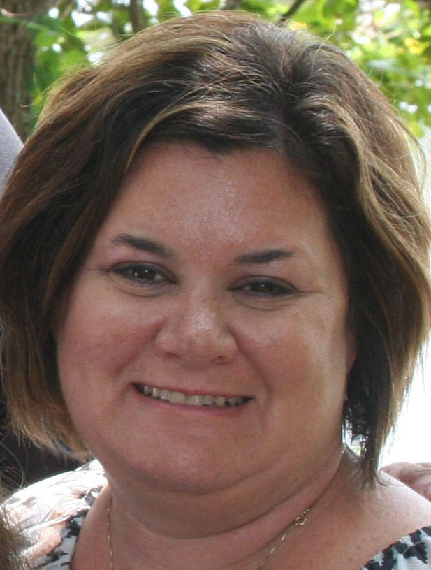
Jennifer G. Elee has been an Environmental Scientist with the Louisiana Department of Environmental Quality for over 20 y. She conducts state inspections of radioactive material licensees, mammography facilities, and x ray registrants. Ms. Elee participates in nuclear power plant exercises and provides support for all radiological emergency response. She provides training for state inspectors and radiologic technologists as needed. Ms. Elee has also been an active member in the Conference of Radiation Control Program Directors for many years and is currently serving on the Board of Directors as a Member-at-Large and is over the Healing Arts Council. |
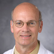
Donald P. Frush is the John Strohbehn Professor of Radiology, and an Associate Faculty Member, Medical Physics Graduate Program at Duke University Medical Center. Dr. Frush earned his undergraduate degree from the University of California Davis, MD from Duke University School of Medicine, was a pediatric resident at University of California San Francisco, completed a radiology residency at Duke Medical Center, and a fellowship in pediatric radiology at Children’s Hospital in Cincinnati. Professional roles included more than 25 y on the Duke Medical Center faculty, with a subsequent nearly 2 y appointment as a Professor of Radiology at Lucile Packard Children’s Hospital at Stanford. He returned to Duke in 2020. Dr. Frush’s research interests are predominantly involved with pediatric body computed tomography (CT), including technology assessment, techniques for pediatric CT examinations, assessment of image quality, radiation dosimetry, and radiation protection and risk communication in medical imaging. Other areas of investigation include CT applications in children and patient safety in radiology. |
JOEL E. GRAY is Professor Emeritus, Mayo Clinic College of Medicine and President of DIQUAD, LLC (Dental Image Quality and Dose), a firm that evaluates dental image quality and dose through the mail. Dr. Gray received his BS in Photographic Science and Instrumentation in 1970, an MS in Optical Sciences in 1974, and a PhD in Radiological Sciences from the Institute of Medical Sciences, University of Toronto in 1977. He served as a Diagnostic Medical Physicist at Mayo Clinic Rochester for 20 y, helped develop and obtain U.S. Food and Drug Administration (FDA) approval for Lorad's (now Hologic) first digital mammography system, and assisted in the development of the microStar patient dosimetry system using optically stimulated dosimetry material while at Landauer, Inc. After leaving Landauer, Dr. Gray founded DIQUAD and continues to operate that business today. Dr. Gray published the first two books on quality control in medical imaging in 1976 under contract to FDA while in graduate school. Dr. Gray is the primary author of the first quality control text (Quality Control in Diagnostic Imaging—A Quality Control Cookbook) which is in use worldwide and has been translated into Chinese. His primary areas of interest include image quality in medical and dental imaging, and optimization of image quality and radiation dose. He serves as a consultant to healthcare organizations and industry. Dr. Gray has served on many national and international advisory committees, including the International Commission Radiological Protection (Committee 3, Radiation Protection in Medicine) and is active in projects with the International Atomic Energy Agency (IAEA) and the World Health Organization. He has co-authored eight publications for the IAEA including educational programs and taught courses for the IAEA in several countries. He has over 170 publications in refereed journals and numerous book chapters, and presented lectures and refresher courses in the United States and overseas. He has visited over 40 countries for both business and pleasure. Dr. Gray was responsible for starting the first Medical Physics Residency Program at Mayo Clinic in 1990. He has mentored masters and doctoral students, and Medical Physics residents. He was elected to NCRP in 1986 and has served on numerous committees producing NCRP Report No. 99, Quality Assurance for Diagnostic Imaging; Report No. 147, Structural Shielding Design for Medical Imaging Facilities; and Report No. 160, Ionizing Radiation Exposure of the Population of the United States. He has served as a Technical Consultant for NCRP Commentary No. 20, Radiation Protection and Measurement Issues Related to Cargo Scanning with Accelerator-Produced High-Energy X Rays; NCRP Report No. 172, Reference Levels and Achievable Doses in Medical and Dental Imaging: Recommendations for the United States; and NCRP Report 177, Radiation Protection in Dentistry and Oral & Maxillofacial Imaging. After serving 18 y on the Council he was named a Distinguished Emeritus Member in 2005. Dr. Gray is a Fellow of the American Association of Physicists in Medicine (AAPM) and the American College of Medical Physics. In 2010 he received the Lifetime Achievement Award from the Upstate New York Association of Medical Physicists and in 2011 the Edith Quimby Lifetime Achievement Award from the AAPM. |
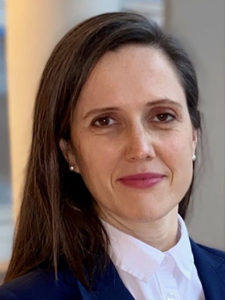
Summer L. Kaplan
is a pediatric radiologist at Children’s Hospital of Philadelphia (CHOP) and Assistant Professor of Clinical Radiology at the University of Pennsylvania Perelman School of Medicine. Dr. Kaplan is the Associate Chair for Quality at CHOP Radiology, is Director for Point-of-Care Ultrasound at CHOP, and is a senior fellow at the Leonard Davis Institute for Health Economics at the University of Pennsylvania. Dr. Kaplan serves on numerous committees with the Radiological Society of North America, American College of Radiology, Society for Pediatric Radiology, and the American Society for Emergency Radiology. She is a member of the Radiation Protection Advisory Committee within the U.S. Department of Environmental Protection in the state of Pennsylvania. Dr. Kaplan’s research includes radiation use in radiography and fluoroscopy as well as emergency and quality improvement topics. Key papers in radiation use include “Quantification of increased patient radiation dose when gonadal shielding is used with automatic exposure control” in the Journal of the American College of Radiology, 2020; “Female gonadal shielding with automatic exposure control increases radiation risks” in Pediatric Radiology, 2018; and “Intussusception reduction: Effect of air vs. liquid enema on radiation dose” in Pediatric Radiology, 2017. Dr. Kaplan has lectured and presented research on these topics at national and international scientific meetings. |

Cari Kitahara
is an epidemiologist and Senior Investigator in the Radiation Epidemiology Branch of the Division of Cancer Epidemiology and Genetics at the National Cancer Institute/National Institutes of Health. She leads a multidisciplinary research program that focuses largely on two, partially overlapping, topics: radiation-associated health risks to medical staff and patients and thyroid cancer etiology and survivorship. She is the Principal Investigator of the U.S. Radiologic Technologists Study (a cohort of 146,000 certified radiologic technologists followed since the mid-1980s), the Cooperative Thyrotoxicosis Therapy Follow-up Study (a cohort of patients treated for hyperthyroidism with radioactive iodine, surgery, or drugs since the 1940s-1960s), and the Pediatric Proton and Photon Therapy Comparison Cohort (a cohort of children treated with either proton or photon radiotherapy and followed over time to assess risks of second cancers – currently recruiting). Her work, including more than 180 peer-reviewed publications and five book chapters, provides high-quality evidence to inform radiation protection practices, cancer prevention efforts, and thyroid cancer clinical care. She is an active member of the American Thyroid Association (ATA) and the International Society for Radiation Epidemiology and Dosimetry (ISoRED), and has recently served on the program and abstract committees of both organizations. She is an Editorial Board member of the journal Thyroid and is the recipient of the prestigious Van Meter Award from the American Thyroid Association and Rosalind Franklin Society Award for Science. |

Emily Marshall
is an Assistant Professor of Radiology and an American Board of Radiology certified Clinical Diagnostic Physicist at the University of Chicago. She serves as a faculty member on the Graduate Program's Committee on Medical Physics. Her research background focused on the development of translational radiation dosimetry models for application in the fluoroscopic imaging of pediatric patient populations. She currently contributes to image quality optimization and clinical testing of all major imaging modalities and maintains primary responsibility for physics support within the Interventional Radiology section. Her ongoing clinical research interests emphasize imaging application optimization in pediatric patient populations, particularly within computed tomography and fluoroscopic applications. |

Sarah McKenney
is a Medical Physicist within the Department of Health Physics and an Associate Professor Volunteer Clinical Faculty within the Department of Radiology at the University of California, Davis (UCD) Medical Center in Sacramento. Dr. McKenney received a BS and BA in Physics and Studio Art from the University of Maryland, College Park. She obtained her PhD in Biomedical Engineering from UCD and completed her medical physics residency at Henry Ford Hospital. Dr. McKenney is a board certified Diagnostic Medical Physicist by the American Board of Radiology. She provides quality assurance for diagnostic imaging systems including general x ray, fluoroscopy, mammography, and computed tomography. Dr. McKenney is Vice Chair of the Pediatric Imaging Subcommittee within the American Association of Physicists in Medicine and she is a steering committee member of Image Gently. Her research interests include imaging quality and dose optimization, particularly for pediatric populations. |
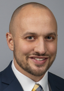
Quentin T. Moore
is a scientific reviewer for the Office of Radiological Health at the Food and Drug Administration’s (FDA) Center for Devices and Radiological Health. He completed his undergraduate education at Mercy College of Northwest Ohio and Washburn University. He earned a MPh from Bowling Green State University and the University of Toledo, and a PhD in Health Science from Northern Illinois University. He is certified by the American Registry of Radiologic Technologists in radiography, radiation therapy, and quality management. Prior to joining FDA, he worked in academia for a decade at Mercy College of Ohio, where he was an Associate Professor and Director of Imaging Sciences. His research focuses on radiation safety culture in medical imaging. Other areas of interest include radiation protection in radiography, and patient safety and quality improvement in medical imaging. He has served on numerous professional committees with organizations such as Image Gently, American Society of Radiologic Technologists, American Association of Physicists in Medicine, and Association of Educators in Imaging and Radiologic Sciences. |

Darcy J. Wolfman
is a Clinical Associate and Clinical Director of Ultrasound in the National Capital Region of the Department of Radiology at Johns Hopkins School of Medicine in Washington, DC. Dr. Wolfman is a board-certified abdominal imager whose expertise is in oncology and genitourinary imaging. Dr. Wolfman is a fellow of the American College of Radiology and the Society of Radiologists in Ultrasound. Dr. Wolfman serves on multiple national committees including the American Association of Physicists in Medicine Ad Hoc Committee on Education and Implementation Efforts for Discontinuing the Use of Patient Gonadal and Fetal Shielding (CARES committee), American College of Radiology Panel on Appropriateness Criteria-Systemic Oncology, and the Ultrasound Online Assessment Committee for the American Board of Radiology. Dr. Wolfman also has been involved with multiple allied health organizations including currently serving as a Trustee on the American Registry of Radiologic Technologists Board of Trustees. |

HELEN A. GROGAN
is President of Cascade Scientific, Inc., an environmental consulting firm. Dr. Grogan received her PhD from Imperial College of Science and Technology at the University of London in 1984 and has more than 25 y of experience in radioecology, environmental dose reconstruction, and the assessment of radioactive and nonradioactive hazardous wastes. She first worked at the Paul Scherrer Institute in Switzerland on the performance assessment of radioactive waste disposal for the Swiss National Cooperative for the Disposal of Radioactive Waste (Nagra). Dr. Grogan was actively involved in the early international cooperative efforts to test models designed to quantify the transfer and accumulation of radionuclides and other trace substances in the environment. Validation of computer models developed to predict the fate and transport of radionuclides in the environment remains a key interest of hers. In 1989 Dr. Grogan returned to the United Kingdom as a senior consultant to Intera Information Technologies before moving to the United States a few years later, where she has worked closely with Risk Assessment Corporation managing the technical aspects of a wide variety of projects that tend to focus on public health risk from environmental exposure to chemicals and radionuclides. Dr. Grogan has served on committees for the National Academy of Sciences, the International Atomic Energy Agency, the U.S. Environment Protection Agency, and NCRP. She co-edited the text book Radiological Risk Assessment and Environmental Analysis published by Oxford University Press in July 2008, and authored the chapter on Model Validation. |


 News & Events